Home » Biomechanical study of the footprint
Biomechanical study of the footprint
Advanced analysis to detect alterations that affect knees, hips and back
Gait analysis is an art as well as a science.
Our Podomèdic Centers are equipped with the latest advances for the study of biomechanics, which allows us to identify possible anomalies related to our way of walking and obtain a better diagnostic orientation for their treatment or prevention, both in a static (standing) and dynamic position. .
What is the biomechanical study of gait or footprint?
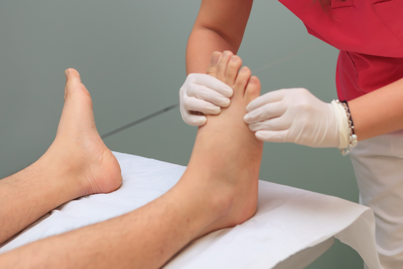
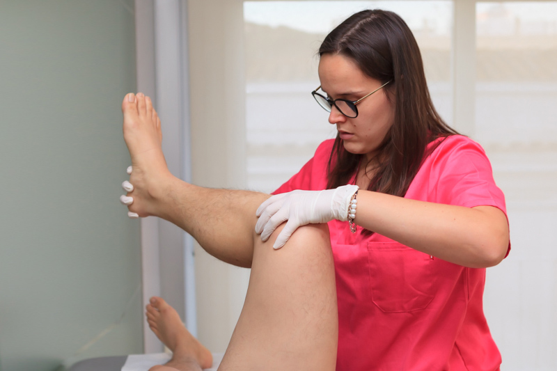
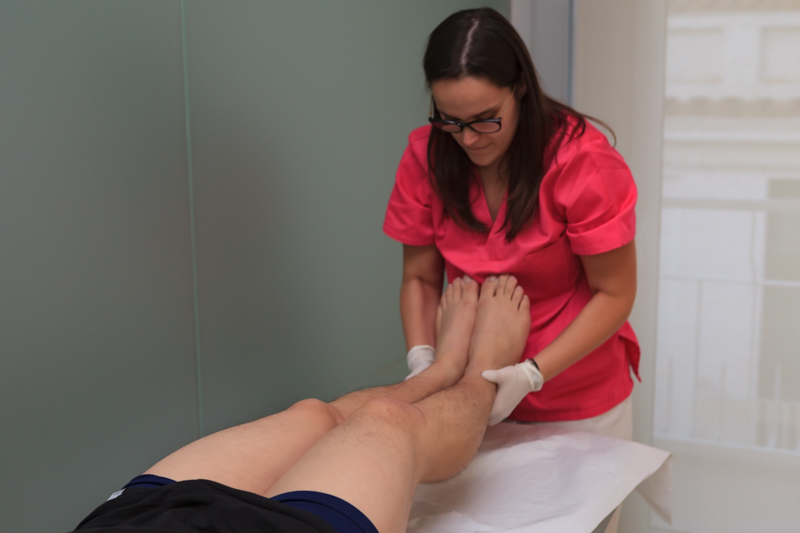
It consists of the analysis of the foot in a static and dynamic position in order to obtain the maximum information on how the muscles, bones and joints of our locomotor system work and where its center of gravity is located.
It should be borne in mind that a large part of ankle, hip and spine injuries are directly related to our way of walking. The foot is the base where the entire weight of the body is supported and if the foundations are poorly supported some parts of our body will suffer.
How do we carry out the biomechanical study in our Podomèdic Centers?
- Study of the tread in statics and dynamics
- Foot scan
- Ultrasound
- Podoscope
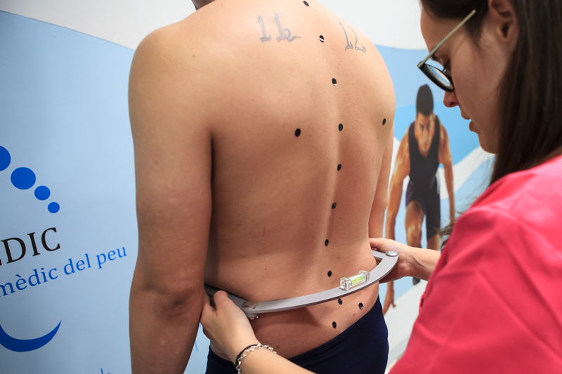
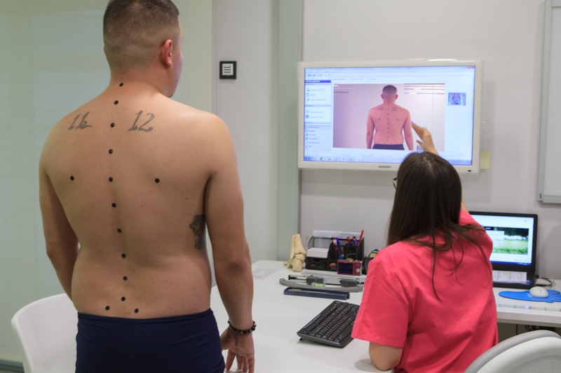
- Spinal scan
- Running scan
- Foot scan in 2D photopogram and 3D molds
Study of the tread in statics and dynamics
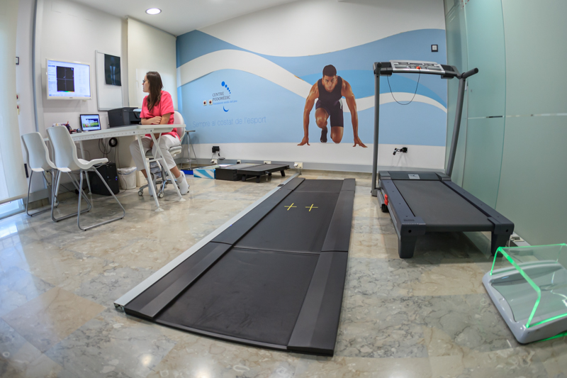
We carry out the study biomechanical tread through a platform of 3 meters that has a digitized system of sensors that detects the plantar pressures both in statics and dynamics.
Through this platform we can know the distribution of the pressures of the plantar footprint and quantify the transmission of loads on the feet and identify the type of foot according to its support, in addition to allowing us to automatically measure the effectiveness of the plantar supports.
Static:
Analyze plantar pressures while the patient is standing.
Dynamic
We make the patient walk several times on the platform, which allows us to detect the distribution of pressures in different areas of the sole of the foot.
Through an electronic record we can see on our computer screen a map of plantar pressures with colors.
It should be taken into account that a large part of ankle, hip, and spine injuries are directly related to our way of walking. The foot is the base where the entire weight of the body is supported, and if the foundations are poorly supported, some parts of our body will suffer.
The biomechanics of the foot, ankle, knee and hip is extremely complex. Therefore, the application of an orthopedic treatment requires a rigorous and precise exploration of the musculoskeletal system.
Foot scan
Ultrasound
Ultrasound scanners are the best tool for to diagnose and treat foot problems and diseases.
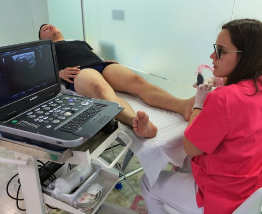
In Podomedic Centers we have ultrasound machines which allow us to have a graphic representation of the feet. We could say that it works like "an eye into the human body." Thanks to the ultrasound we can assess from the first moment the state in which the damaged structure is, monitor the healing process and assess whether the treatment designed individually for each patient is being effective.
- Ultrasound allows us explore various structures of the musculoskeletal apparatus, such as muscles, tendons, ligaments, bursae (cavities filled with fluid), cartilage and bone surfaces.
- In addition, another of the attributes of the ultrasound machine is that it makes it possible establish a protocol acting and follow-up to assess the evolution of an injury.
- And it is also useful for perform ultrasound-guided infiltrations. This entails being able to inject the desired substance at the exact point of the ailment.
It is a non-invasive work tool that allows you to see the image of the lesion in real time and make an instant diagnosis.
The most frequent pathologies are:
- Ligamentous pathology: ankle sprains
- Tendon pathology: tenosynovitis, Achilles tendon pathology, intratendinous calcifications, and so on
- Muscle pathology: partial breakage
- Bone pathology: tibial stress, stress fracture
- Morton's neuroma
- Plantar plate rupture
- Plantar fasciitis
- Cysts
Podoscope
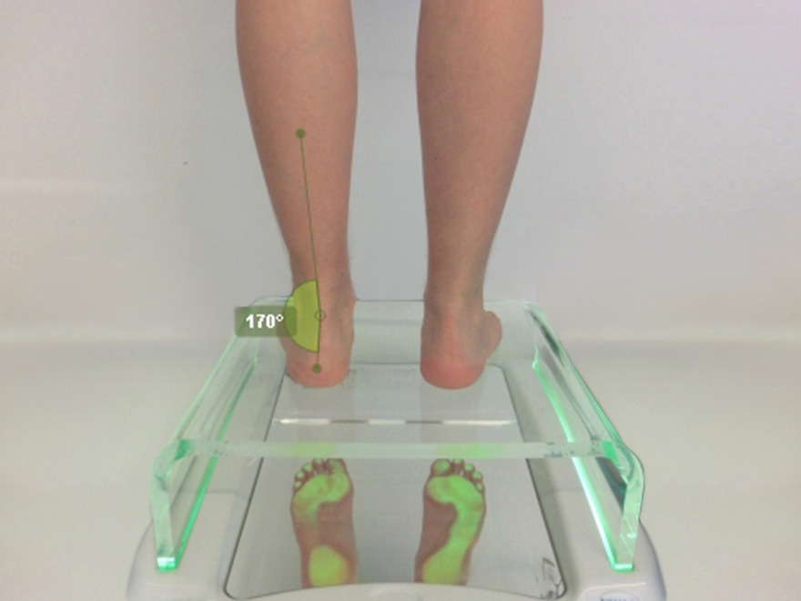
Examination while standing (standing) using a podoscope. It allows us to quickly and safely visualize and study the plantar footprints, the different axes of the feet and check the applied treatments.
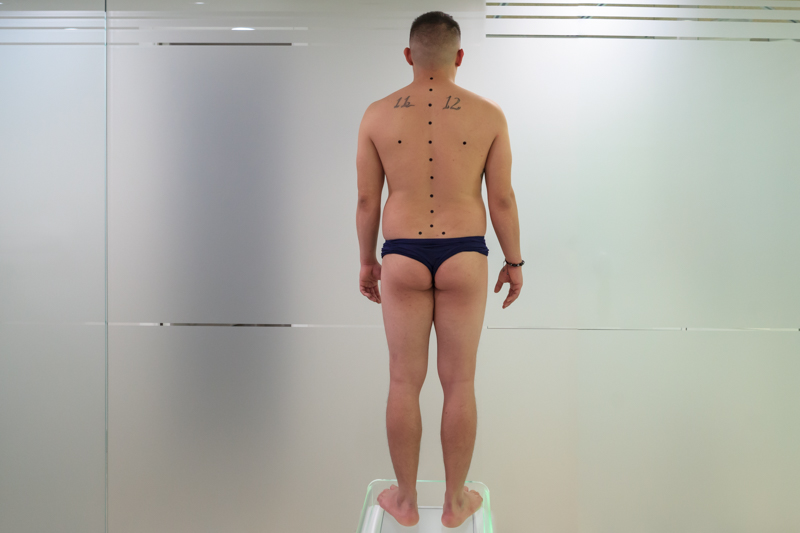
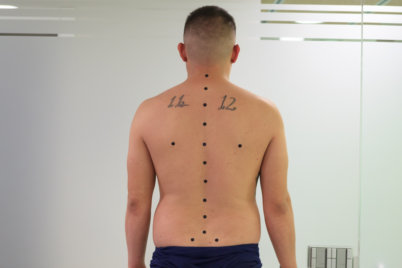
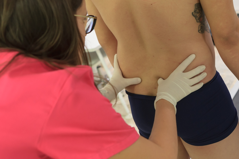
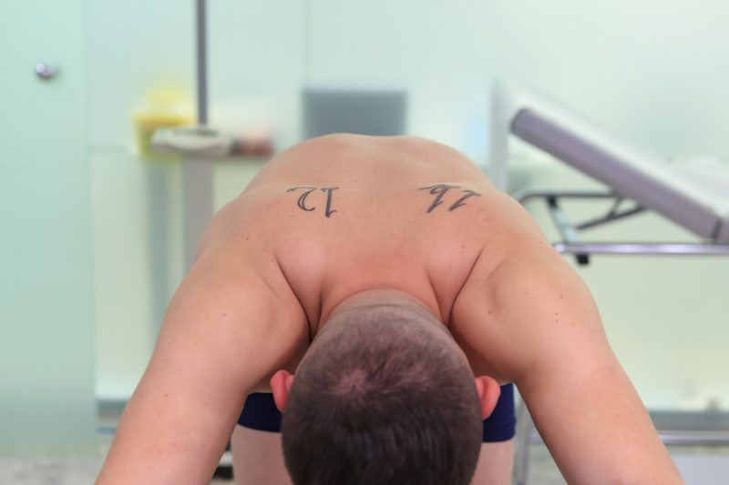
Spinal scan
Treadmill scan
It allows us to film the patient in order to make a complete assessment of the footprint while running or walking on the treadmill.
2D and 3D foot scan
2D scanner:
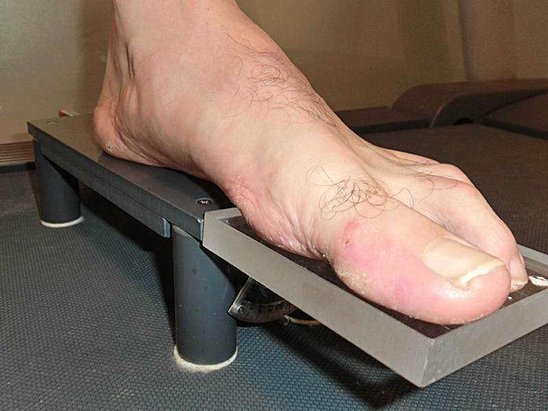
The 2D scanner gives us information on the patient's footprint, specifically, the overload areas, the contour of the plantar imprint and the exact number of the foot (among other parameters).


3D scanner:
In Podomedic Centers we use the 3d scanner of the latest generation that guarantees a more efficient, fast and precise design of the custom orthotics.


Thanks to this great advance, we obtain a three-dimensional footprint. This system allows us to make templates with state-of-the-art technology, since it sends the scanner data to a computerized milling machine that makes the custom template (orthosis) with the highest quality standards.
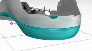
In addition, they allow us to rectify, during the scan, the incorrect position of the foot and once the mold is obtained on the computer screen, make the appropriate modifications.