What is cavus foot?
Cavus foot is an anatomical malformation in which the foot arch is higher than normal. This involves a modification of soil support and a different distribution of body weight at the feet. People who do not present any pathology at the feet redistribute their body weight into three specific parts of the foot; in the heel, the outer central and front (the fingers), instead, people who suffer from cavus foot spread all weight in the heel and in the front (metatars) area.
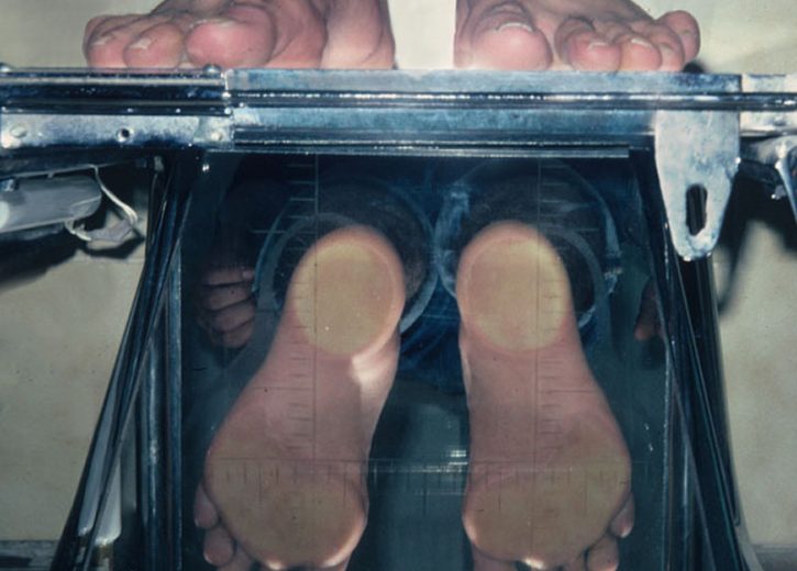


It should be noted that cavus foot can be asymptomatic. However, when cavus foot is symptomatic, signs may include:
- Pain or unease at the feet, especially on the sides or in the metatarsal area.
- Pain and instability in ankles, which predicts repeated squinches.
- Excessive development of the longitudinal arch of the foot accompanied by the shortening of the extending muscles of the fingers, giving rise to pathologies such as hammertoe or clawtoe.
- Elevation of the plant and retraction of the fascia causing the foot to be shortened.
- Callosity parking in areas of maximum pressure, such as the heel, or the metatarsal zone.
- Difficulty standing for many hours at a time, walking long distances, or running because the pain sensation is intensified.
Listening to the patient, performing a good physical examination and conducting an analysis of his medical history is usually a sufficient protocol for obtaining correct diagnosis and planning the most appropriate treatment.
We say listening to the patient is important because the standing diagnosis of cavus foot includes a review of family background as it is usually a hereditary condition.
We carry out a biomechanical study of the footprint and examine whether the patient has any neurological alteration. If the high arch of the foot is due to a neurological disorder or another medical condition, it is likely to get progressively worse.
For this reason, we often ask for other diagnostic tests, including X-rays, nuclear magnetic resonance (RMN) of brain and spinal cord and electromyography.
They can be of congenital or acquired origin.
Most people with cavus foot have a neurological disorder, which may be static (symptoms do not change) or progressive (symptoms become worse over time).
Static neurological disorders that cause cavus foot include: stroke, polio, cerebral palsy, spinal injury, and peroneal nerve injury.
Progressive neurological disorders that can cause cavus foot include spinal tumor, brain tumor, spinal cyst, muscular dystrophy, Friedreich ataxia, and Charcot-Marie-Tooth syndrome.
Depending on the severity of the symptoms, treatment can be conservative or surgical.
Conservative treatment of the cavus foot may include one or more of the following options:
Chiropody. In case the patient presents harshness or corns in the support areas.
Orthopedic devices. Customized insoles that fit the shoe and are beneficial because they provide stability and cushioning to the foot, increasing the contact surface and improving load distribution.
Silicone digital orthosis. To protect the fingers that are in digital harp.
Footwear modifications. The shoes holding the ankle and the shoes with broad heels give stability.
Physiotherapy exercises. To improve the ankle muscles and achieve greater stability. They may be accompanied by stretching exercises (or muscle lengthening) in order to strengthen all muscles in the leg.
If conservative treatment fails to relieve pain and improve foot stability, surgery may be needed.